寄生蠕虫類の分類体系.第Ⅲ巻.脊椎動物の線虫類 原図 1/10
きせいぜんちゅうるいのぶんるいたいけい.だいさんかん.せきついどうぶつのせんちゅうるい げんず じゅうぶんのいち
概要
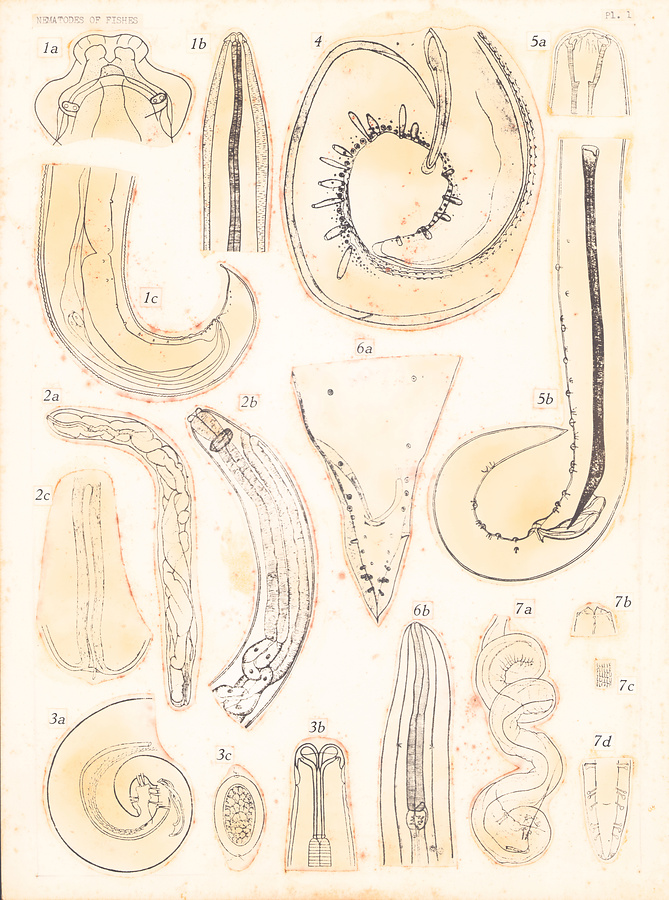
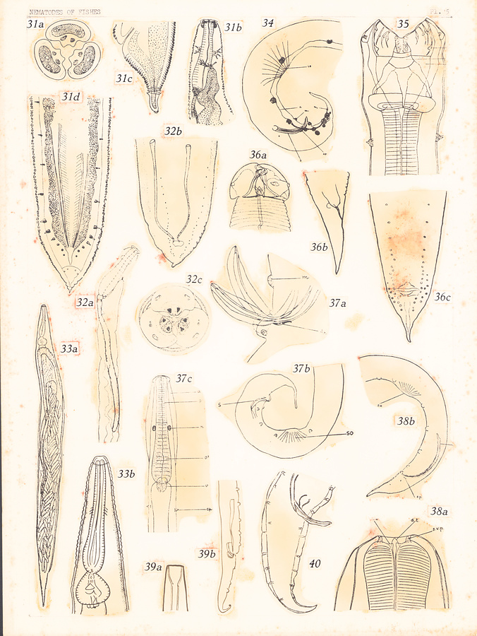
Plate 1.
Fig. 1. Raphidascaris acus (Bloch, 1779). After Mozgovoi, 1951.a. Head, dorsal view. b. Anterior extremity of male. c. Posterior extremity of male.
Fig. 2. Cystoopsis acipenseris Wagner, 1867. After Saidov, 1954. a. Entire female. b. Anterior extremity of female. c. Posterior extremity of male.
Fig. 3. Metabronema magnum (Taylor, 1925). After Yorke and Maplestone, 1926. a. Posterior extremity of male. b. Anterior extremity of male. c. Egg.
Fig. 4. Comephoronema werestschagini Layman, 1933; posterior extremity of male.
Fig. 5. Filochona sulaki Saidov, 1954.
Anterior extremity of male.
Posterior extremity of male.
Fig. 6. Cottocomephoronema problematicum Layman, 1933. a. Posterior extremity of male. b. Anterior extremity of male.
Fig. 7. Heliconema heliconema Travassos, 1919. a. Spiral posterior extremity of male. b. Head of male, ventral view. c. Cuticle showing serration. d. Posterior extremity, ventral view.
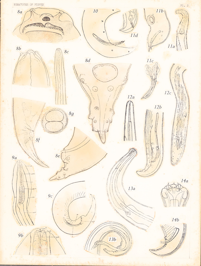
Plate 2.
Fig. 8. Pseudanisakis rotundata (Rud., 1819). After Mozgovoi, 1950.
a. Head. b—c. Anterior extremity of male. d—e. Posteriorextremity of male. f. Posterior extremity of female. g. Egg.
Fig. 9. Paraquimperia tenerrima (Linstow, 1878). After Baylis, 1934. a—b. Anterior extremity of male. c. Posterior extremity of male.
Fig. 10. Spironoura leptocephala Baylis et Daubney, 1922; posterior extremity of male.
Fig. 11. Spectatus spectatus (Travassos, 1923). After Travassos, 1928. a. Anterior extremity, lateral view. b. Posterior extremity of male. c. Spicules and gubernaculum. d. Posterior extremity of female.
Fig. 12. Sterliadochona ssavini Skrj., 1946. After Skrjabin, 1949.
a. Anterior extremity of male. b—c. Posterior extremity of male.
Fig. 13. Haplonema immutatum Ward et Magath, 1917, In Skrjabin et al., 1949. a. Anterior extremity of male. b. Posterior extremity of male.
Fig. 14. Nematoxynema piscicola (Linstow, 1907). a. Anterior extremity of male. b. Posterior extremity of male.
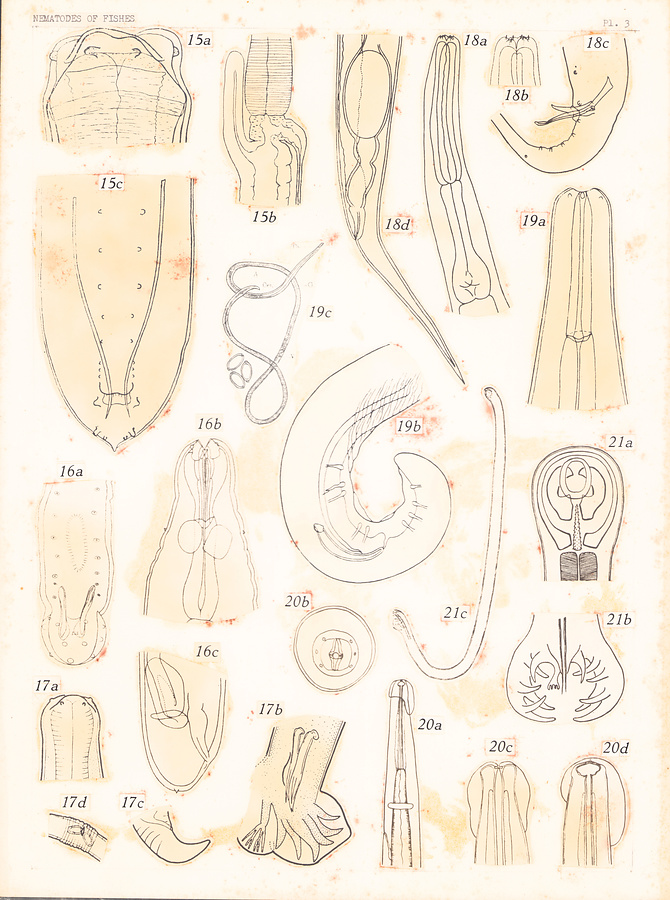
Plate 3.
Fig. 15. Heterotyphlum himantolophi Spaul, 1927. a. Anterior extremity of male. b. Ventricular appendix and intestinal cecum of male. c. Posterior extremity of male.
Fig. 16. Neocucullanus neocucullanus Travassos, Artigas et Pereira, 1928. a. Posterior extremity of male. b. Anterior extremity of male. c. Posterior extremity of female.
Fig. 17. Ichthyostrongylus clelandi Mawson, 1954. a. Anterior extremity of male. b. Posterior extremity of male. c. Posterior extremity of female. d. Vulvar region of female.
Fig. 18. Rondonia rondoni Travassos, 1919. After Baylis, 1936, in Skrjabin et al., 1951. a—b. Anterior extremity of male. c. Posterior extremity of male. d. Posterior extremity of female.
Fig. 19. Capillospirura ovotrichuria Skrjabin, 1924. After Ivanov and Murygin, 1937. a. Anterior extremity of male. b. Posterior extremity of male. c. Female and eggs.
Fig. 20. Parascarophis sphyrnae Campana-Rouget, 1955. a. Anterior extremity of male. b. Head of male, end-on view. c. Head of male, dorsoventral view. d. Same, lateral view.
Fig. 21. Cylicostrongylus ciureai Dinulescu, 1943. a. Head, dorsal view. b. Bursa. c. Male.
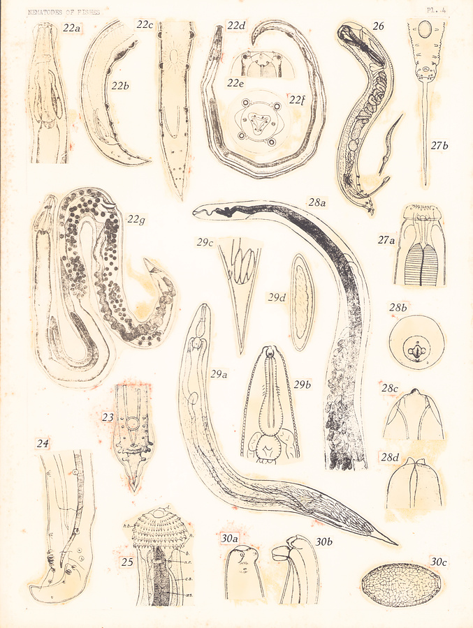
Plate 4.
Fig. 22. Buckleynema buckleyi Ali et Singh, 1954. a. Anterior extremity of male, lateral view. b. Posterior extremity of male, lateral view. c. Same, ventral view. d. Male. e. Head of male, lateral view. f. Same, end-on view. g. Female.
Fig. 23. Ascaridia brevicauda (Rátz, 1897); posterior extremity of male.
Fig. 24. Echinocephalus spinossimus Shipley et Hornell, 1905; posterior extremity of male. After Baylis and Lane in Baylis, 1939.
Fig. 25. Echinocephalus uncinatus Molin, 1858; anterior extremity of male. After Baylis and Lane, 1920.
Fig. 26. Cucullanellus dichelyneformis Szidat, 1950; male.
Fig. 27. Kathlania chiloscyllii Thwaite, 1927. a. Anterior extremity of male. b. Posterior extremity of male.
Fig. 28. Comephoronema werestchagini Layman, 1933. a. Anterior extremity of female. b. Head, end-on view. c. Same, lateral view. d. Same, dorsoventral view.
Fig. 29. Cosmoxynema vianai Travassos, 1949. a. Female. b. Anterior extremity of female. c. Posterior extremity of female. d. Egg.
Fig. 30. Capillaria baicalensis Rushkov et Sudarikov, 1954. a—b. Posterior extremity c. Egg.
Plate 5.
Fig. 31. Goezia (Pseudogoezia) sigalasi Stefanski, 1938. a. Head, end-on view. b. Anterior extremity of male. c. Posterior extremity of female. d. Posterior extremity of male.
Fig. 32. Goezia (Goezia) spinulosa (Dies.). After Freitas and Lent, 1946. a. Anterior extremity of male. b. Posterior extremity of male. c. Head, end-on view.
Fig. 33. Cosmoxynemoides aguirrei Travassos, 1949. a. Female. b. Anterior extremity of female.
Fig. 34. Metaquimperia bagarii Karve, 1941; posterior extremity of male.
Fig. 35. Paracamallanus ophiocephali Karve, 1941; anterior extremity of male.
Fig. 36. Contracaecum iheringascaris (Pereira, 1935). a. Head of male. b. Posterior extremity of female. c. Posterior extremity of male.
Fig. 37. Metaquimperia callichroi Karve, 1941. a. Spicules of male. b. Posterior extremity of male. c. Anterior extremity of male.
Fig. 38. Gendria tilapiae Baylis, 1930. a. Head of male. b. Posterior extremity of male.
Fig. 39. Johnstonmawsonia coelorhynchi (Johnston et Mawson, 1945). After Johnston & Mawson, 1945. a. Head of male. b. Posterior extremity of male.
Fig. 40. Rhabdochona beaufortis (Liu et Wu, 1941); posterior extremity of male.
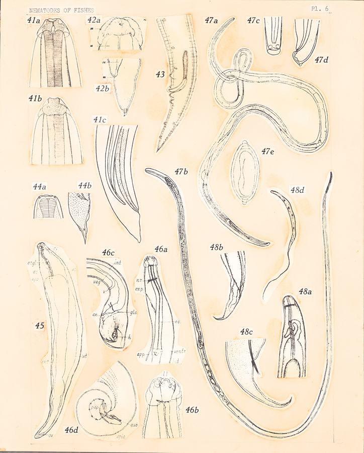
Plate 6.
Fig. 41. Raphidascaris acus (Bloch, 1779). After Yorke and Maplestone, 1926. a. Head, ventral view. b. Head, dorsal view. c. Posterior extremity of male.
Fig. 42. Neogoezia magna Kreis, 1937. a. Head. b. Posterior extremity of female.
Fig. 43. Ichthyobronema conoura (Linstow, 1885); posterior extremity of male.
Fig. 44. Paraseuratum tandani Johnston et Mawson, 1940. a. Anterior extremity of male. b. Posterior extremity of male.
Fig. 45. Clavinema parasiluri Yamaguti, 1935; female.
Fig. 46. Hedruris bryttosi Yamaguti, 1935. a. Anterior extremity of male. b. Head, lateral view. c. Posterior extremity of female. d. Posterior extremity of male.
Fig. 47. Capillaria zederi (Teixeira de Freitas et Lent, 1935). a. Male. b. Female. c—d. Posterior extremity of male. e. Egg.
Fig. 48. Philonema oncorhynchi Kuitun.-Ekb., 1933. a. Anterior extremity of female. b. Posterior extremity of female. c. Posterior extremity d. Embryo.
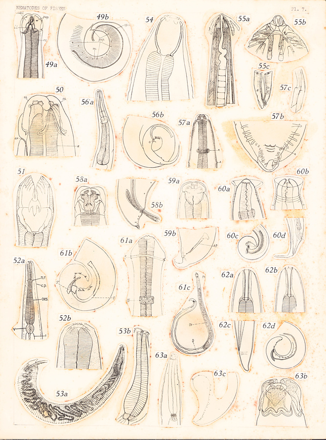
Plate 7.
Fig. 49. Cotvacanecum pagvosomi Yamaguti, 1935. a. Anterior extremity of male. b. Posterior extremity of male.
Fig. 50. Paranisakiopsis coelorhynchi Yamaguti, 1941; anterior extremity of female.
Fig. 51. Pavacamallanus cyathopharynx (Baylis, 1923); anterior extremity of male.
Fig. 52. Acanthocheilus quadridentatus Molin, 1858. After Baylis, 1929. a. Anterior extremity of male. b. Head of male.
Fig. 53. Goezia ascaroides (Goeze, 1782). After Goeze in Yorke and Maplestone, 1926. a. Male. b. Anterior extremity of male.
Fig. 54. Procamallanus laeviconchus (Wedl, 1861 After Yorke and Maplestone, 1926.
Fig. 55. Ancyracanthus pectinatus Dies., 1838. After Yorke and Maplestone, 1926. a. Anterior extremity of male. b. Head of same. c. Posterior extremity of male.
Fig. 56. Monhystevides piscicola Baylis et Daubney, 1922. a. Anterior extremity of male. b. Posterior extremity of male.
Fig. 57. Porrocaecum draschei (Stoss., 1896). After• Baylis, 1927. a. Anterior extremity, dorsal view. b. Posterior extremity of male. c. Spicules.
Fig. 58. Paranisakis squatinae Baylis, 1923. a. Anterior extremity of male. b. Posterior extremity of male.
Fig. 59. Dujardinascaris malapteruri Baylis, 1923. a. Anterior extremity of male. b. Posterior extremity of male.
Fig. 60. Proleptus obtusus Duj., 1845. After Yorke and Maplestone, 1926. a—b. Anterior extremity of female. c. Posterior extremity of male. d. Posterior extremity of female.
Fig. 61. Cyclozone acipenserina Dogiel, 1932. a. Anterior extremity of male. b. Posterior extremity of male. c. Male.
Fig. 62. Cystidicola farionis Fischer, 1798. After Yorke and Maplestone, 1926. a. Anterior extremity of female. b. Same of male. c. Posterior extremity of female. d. Posterior extremity of male.
Fig. 63. Ichthyanisakis monodi Gendre, 1928. a. Anterior extremity of male. b. Head of male. c. Posterior extremity of male.
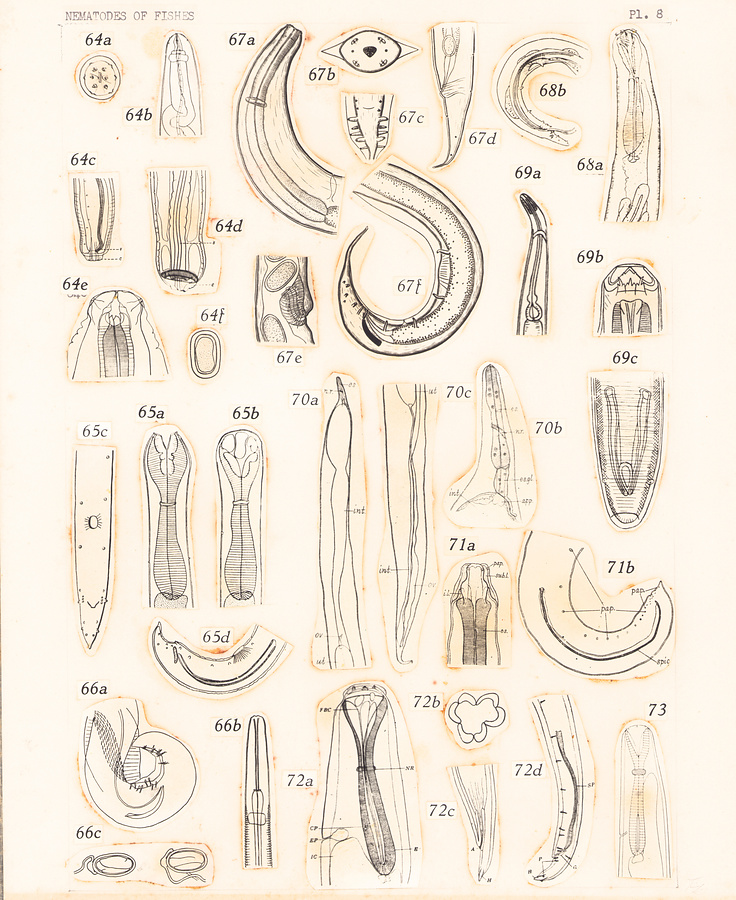
Plate 8.
Fig. 64. Eustrongylides wenrichi Canavan, 1929. a. Head, end-on view. b. Anterior extremity of male, lateral view. c. Posterior extremity of female. d. Same of male. e. Anterior extremity of male, ventral view. f. Egg.
Fig. 65. a. Anterior extremity of Cucullanus carettae Baylis, 1923, ventral view. After Y. & M., 1926. b. Same, lateral view. After Y. & M., 1926. c. Posterior extremity of male of Cucullanus cirratus Müller, After Schneider in Y. & M., 1926. 1777. d. Posterior extremity of male of Cucullanus carettae Baylis, After Y. & M., 1926. 1923.
Fig. 66. Ascarofrhis morrhuae Beneden, 1871. After Gordon, 1951. a. Posterior extremity of male. b. Anterior extremity of male. c. Eggs.
Fig. 67. Pingus sinensis Hsü, 1933. a. Anterior extremity, lateral view. b. Head, end-on view. c. Posterior extremity of male. d. Posterior extremity of female. e. Vulva. f. Posterior extremity of male.
Fig. 68. Dacnitoides cotylophora Ward et Magath, 1917. a. Anterior extremity of female. b. Posterior extremity of male.
Fig. 69. A mblyonema terdentatum Linstow, 1898. After Linstow in Yorke and Maplestone, 1926. a. Anterior extremity of male. b. Head. c. Posterior extremity of male.
Fig. 70. Ichthyofilaria dasycotti Yamaguti, 1935. a—b. Anterior extremity of female. c. Posterior extremity of female.
Fig. 71. Orantoisakis lophii (Yamaguti, 1935). a. Anterior extremity of male. b. Posterior extremity of male.
Fig. 72. Neocucullanellus apharei Yamaguti, 1941. a. Anterior extremity, lateral view. b. Transverse section of false buccal capsule, c. Posterior extremity of female. d. Posterior extremity of male.
Fig. 73. Dichelyne fossor Jägerskiöld, 1902; anterior extremity, lateral view.
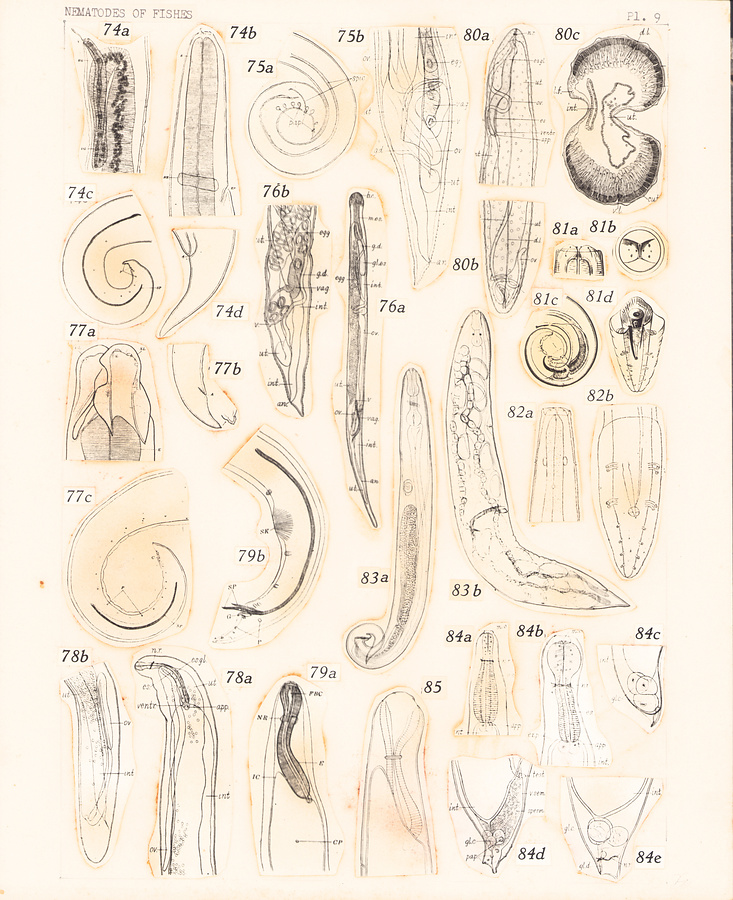
Plate 9.
Fig. 74. Metanisahis vaiae (Yamaguti, 1941). a. Female terminalia. b. Anterior extremity, lateral view. c. Posterior extremity of male. d. Posterior extremity of female.
Fig. 75. Spinitectus gigi Fujita, 1928. a. Posterior extremity of male. b. Posterior extremity of female.
Fig. 76. Camallanus cotti Fujita, 1927. a. Female. b. Posterior extremity of female.
Fig. 77. Raphidascaroides nipponensis Yamaguti, 1941. a. Head, ventrolateral view. b. Posterior extremity of female. c. Posterior extremity of male.
Fig. 78. Philometra lateolabracis (Yamaguti, 1935). a. Anterior extremity of female. b. Posterior extremity of female.
Fig. 79. Cucullanellus branchiostegi Yamaguti, 1941. a. Anterior extremity, lateral view. b. Posterior extremity of male.
Fig. 80. Philometroides seriolae (Ishii, 1931). After Yamaguti, 1936. a. Anterior extremity of female. b. Posterior extremity of female. c—d. Transverse section of female through uterus and intestine.
Fig. 81. Heliconema anguillae Yamaguti, 1933 = Ortleppina longissima (Ortlepp, 1922) of Li, 1934. After Li, 1934. a. Head, ventral view. b. Same, end-on view. c—d. Posterior extremity of male.
Fig. 82. Physaloptera longissima (Ortlepp, 1922). After Ortlepp, 1922. a. Anterior extremity of male. b. Posterior extremity of male.
Fig. 83. Travnema travnema Pereira, 1938. a. Male. b. Female.
Fig. 84. Anguillicola globiceps Yamaguti, 1935. a. Anterior extremity of larva. b. Same of male, lateral view. c. Posterior extremity of larva. d. Same of male, lateral view. e. Same of female, lateral view.
Fig. 85. Cucullanellus minutus (Rud., 1819); anterior extremity, lateral view. After Törnquist, 1931.
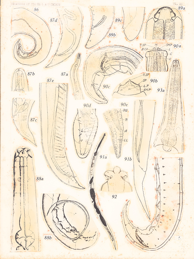
Plate 10.
Fig. 86. Foleyella americana Walton, 1939; posterior extremity of male.
Fig. 87. Cruzia morleyi (Pearse, 1936). a. Anterior extremity of male. b. Head. c. Posterior extremity of female. d—e. Posterior extremity of male.
Fig. 88. Dibulbiger longispiculis Caballero, 1935. a. Anterior extremity of male. b. Posterior extremity of male.
Fig. 89. Amplicaecum brumpti Khalil, 1926. a. Head, dorsal view. a. Posterior extremity of male. b. Posterior extremity of female.
Fig. 90. Paraleptus scyllii Wu, 1927, a. Anterior extremity, ventral view.
b. Mouth lip, lateral view. c—d. Posterior extremity of male. e. Posterior extremity of female.
Fig. 91. Raillietnema simplex (Travassos, 1925). a. Female. b. Posterior extremity of male.
Fig. 92. Spiroxys japonica Morishita, 1926; head, lateral view
Fig. 93. Paracosmocerca mucronata Kung et Wu, 1954. a. Anterior extremity of male. b. Posterior extremity of male.
所蔵館のウェブサイトで見る
公益財団法人 目黒寄生虫館文化庁 〒602-8959 京都府京都市上京区下長者町通新町西入藪之内町85番4
(C) The Agency for Cultural Affairs